

close
Choose Your Site
Global
Social Media
Views: 0 Author: Site Editor Publish Time: 2026-06-24 Origin: Site


Modern clinical practice increasingly prioritizes minimally invasive interventions over traditional large-bore procedures. Healthcare providers now actively seek advanced systems that prioritize patient tolerance alongside optimal clinical outcomes. Unfortunately, standard medical tubing frequently faces significant failure points in daily practice. Premature migration, frequent occlusion, and severe patient discomfort often complicate prolonged retention scenarios. These pressing challenges drive the urgent demand for superior, dependable alternatives. You can rely on the double pigtail drainage tube as a highly effective standard-of-care solution. It masterfully balances secure internal anchoring against efficient, continuous fluid evacuation. This versatile performance makes it ideal across diverse anatomical environments, including ureteral, biliary, and pleural spaces. In this comprehensive guide, you will explore the precise biomechanics, advanced material science, and crucial clinical nuances defining these devices. We will detail unique retention mechanisms, compare performance profiles against traditional options, and outline practical implementation protocols.
Dual-Anchoring Mechanism: The bilateral coiled design minimizes migration risks at both the proximal and distal ends without requiring external suturing or balloon inflation.
Material Dependability: Modern iterations utilizing specialized polyurethane offer a high inner-to-outer diameter ratio, maximizing flow in a smaller footprint.
Clinical Equivalence: Data consistently supports small-bore pigtail catheters as safe, effective alternatives to traditional large-bore chest tubes for specific fluid and air evacuations.
Procurement Priority: Evaluating these tubes requires strict attention to radiopacity, kink resistance, and biocompatible coatings to reduce encrustation.
How does an implanted device stay exactly where you put it? The fundamental secret lies in structural memory. Modern polymers undergo specific thermal setting during manufacturing. This process trains the coiled ends to naturally curl back into their original shape immediately after insertion. This unique mechanical memory allows the tube to securely anchor within confined, irregular cavities. Common clinical applications include the renal pelvis or the contracting bladder. You do not have to worry about spontaneous dislodgement as the patient moves. Furthermore, this elegant design completely eliminates the need for traumatic anchoring mechanisms. Rigid plastic flanges and inflatable balloons often cause severe mucosal irritation over time. Balloons can deflate unexpectedly or cause pressure necrosis. The coiled tips provide a soft, atraumatic bumper instead. They gently rest against delicate tissues, absorbing minor shifts without causing harm.
Effective fluid evacuation requires much more than a single open tip. Look closely at the inner curve of these specialized coils. You will find multiple precise fenestrations, commonly known as side holes, carefully distributed along the loop. This multi-port configuration drastically improves overall drainage efficiency. If one hole becomes temporarily blocked by floating debris, the others continue functioning seamlessly. The coiled structure itself serves a crucial dual purpose here. It physically suspends the tube and pushes sensitive mucosal tissue away from the active drainage holes. Consequently, the design prevents surrounding tissue from being aggressively aspirated into the lumen. Continuous patency remains remarkably stable, even during active, applied suction. This spatial separation guarantees that the vacuum forces only act upon the target fluid.
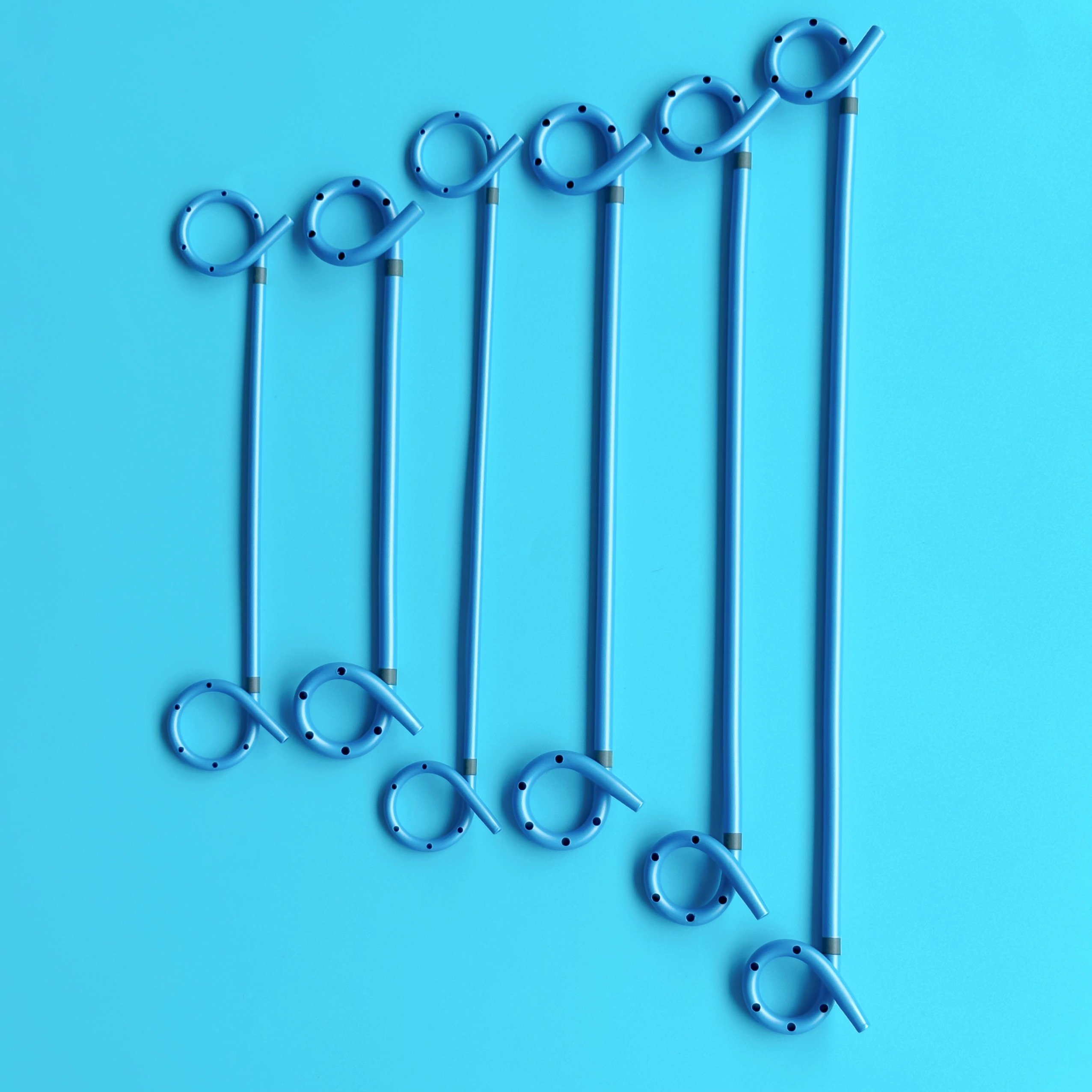
Clinical teams frequently compare insertion profiles when selecting vital hospital equipment. The pigtail drainage tube offers a distinctly superior advantage regarding tissue trauma. Providers typically insert these devices using the well-established Seldinger technique. This guidewire-assisted micro-insertion method requires only a tiny, precise incision. An operator sequentially dilates the tract over a wire, minimizing collateral damage. Conversely, traditional large-bore straight tubes often demand aggressive blunt dissection of multiple muscle layers. This stark procedural difference profoundly impacts the patient experience. Smaller incisions translate directly to significantly lower postoperative pain scores. Patients also regain their physical mobility much faster. They tolerate the compact profile exceptionally well during prolonged recovery periods, facilitating earlier hospital discharge.
We must clearly define where these small-bore devices fail. Recognizing limitations builds essential clinical trust. No single surgical tool fits every complex scenario perfectly. A standard medical drainage tube with a small French size struggles against highly viscous fluids. Straight, large-bore chest tubes remain absolutely necessary for specific severe conditions. For example, heavy hemothorax or thick, organizing empyema demands massive internal lumens. If you deploy a small 8-French pigtail for thick pus or heavy blood clots, it will inevitably suffer rapid occlusion. Fibrin easily bridges the tiny fenestrations, rendering the device useless. Always evaluate the specific fluid properties meticulously before selecting a small-bore device. Appropriate patient selection prevents preventable emergency exchanges.
Material selection directly governs how a plastic drainage catheter behaves deep inside the human body. Polyurethane currently dominates the modern double pigtail market. It possesses remarkably high tensile strength at a molecular level. This structural strength allows manufacturers to engineer much thinner walls. Thinner walls directly translate to larger internal lumens and vastly higher fluid flow rates. Polyurethane also provides excellent, reliable kink resistance during complex bending. Silicone, alternatively, offers undeniably superior biocompatibility. It feels considerably softer and triggers less localized inflammatory reactivity during multi-month placements. However, silicone fundamentally requires much thicker walls to maintain basic structural integrity. This thick-wall requirement inherently reduces the internal flow rate for any given external French size.
Material Attribute | Polyurethane Catheters | Silicone Catheters |
|---|---|---|
Tensile Strength | High; easily withstands rigorous uncoiling and guidewire manipulation | Moderate; requires highly careful handling to avoid tears |
Wall Thickness | Thin; strategically maximizes internal lumen size for fluid flow | Thick; inherently reduces the available internal flow space |
Kink Resistance | Excellent; safely holds internal shape well in tight anatomical angles | Good; but the significantly softer profile can compress easily |
Biocompatibility | Good; highly suitable for standard indwelling durations | Superior; remains the ideal choice for long-term clinical retention |
Bodily fluids continuously interact aggressively with implanted foreign materials. Rapid biofilm formation and crystalline calcification represent harsh realities in urological and biliary tracts. Urine, for instance, quickly deposits hard minerals on untreated polymer surfaces. Advanced surface coatings help extend safe indwelling times significantly. Hydrophilic coatings actively absorb surrounding water molecules. This absorption creates a highly slippery, low-friction fluid boundary layer. This microscopic layer limits dangerous protein adhesion and delays initial bacterial colonization. Some specialized devices also incorporate active antimicrobial layers. These specific technologies aggressively repel harmful pathogens. Utilizing coated devices significantly delays heavy encrustation before a mandatory surgical exchange becomes absolutely necessary.
You cannot safely manage what you cannot see. Precise visualization remains entirely non-negotiable during initial insertion and routine follow-up checks. High-quality devices compound heavy metals like barium, bismuth, or tungsten directly into the polymer matrix. This sophisticated compounding ensures distinct, sharp visibility under active fluoroscopy or standard X-ray imaging. Clear radiopacity allows clinicians to confirm correct anatomical positioning instantly. Furthermore, distinct depth markers uniformly printed along the outer shaft provide vital visual cues. They directly help the operator gauge exact insertion depth at the external skin level. Proper depth verification prevents accidental internal perforation.
Safety during initial placement relies heavily on absolute mechanical integrity. Operators must forcefully straighten the inherently coiled tips over a stiff metallic guidewire prior to insertion. The plastic polymer must easily withstand this severe straightening process without microscopically cracking. Once deployed into the target cavity, it must promptly and reliably uncoil. Structural integrity also matters immensely during routine tube exchanges. If the material degrades from bodily fluid exposure, the catheter might catastrophically fracture during extraction. High tensile strength actively prevents these devastating mechanical failures. It ensures the entire device remains fully intact during removal.
Sourcing standalone tubes often severely complicates fast-paced clinical workflows. Comprehensive, pre-packaged insertion kits elegantly streamline the entire procedural process. Utilizing complete kits aggressively reduces dangerous cross-vendor compatibility issues. Clinical procurement teams must prioritize vendors who supply fully integrated systems. Essential kit components typically include several standardized items:
Compatible Access Needles: Precision echogenic needles designed for clear ultrasound visibility during the initial cavity puncture.
Kink-Resistant Guidewires: Flexible nitinol guidewires that navigate complex anatomical tracts without buckling under pressure.
Graduated Fascial Dilators: Sequentially sized rigid dilators to safely prepare the tissue tract for the final catheter deployment.
Locking Hub Mechanisms: Secure external connection points to attach the fluid line firmly to external collection bags, preventing accidental disconnection.
Providing these tools in one sterile package significantly reduces preparation time. It also firmly guarantees that the wire diameter perfectly matches the internal catheter lumen.
Even the most advanced materials require highly proactive management. Routine flushing protocols often prove absolutely necessary to maintain long-term patency. This applies especially forcefully to complex biliary structures or thick abscess cavities. You must systematically monitor fluid viscosity and output volume closely. A sudden, unexpected drop in drainage output might indicate a severely blocked lumen rather than an empty anatomical cavity. Early, decisive intervention can save the device. Gentle sterile saline flushes often clear minor cellular debris successfully. Always strictly follow established institutional protocols for the specific anatomical space being drained.
Leaving any implanted device in place too long invites serious clinical complications. Aggressive tissue ingrowth occurs frequently if retention exceeds the manufacturer’s recommended duration. Strict tracking of insertion dates across hospital departments reliably prevents this oversight. When physically removing the device, follow standard clinical steps incredibly carefully.
First, securely unlock any external hub locking mechanisms that hold the tension string.
Second, cautiously introduce a flexible, soft-tipped guidewire into the central lumen to gently uncoil the distal tip inside the body.
Finally, smoothly retract the fully straightened device out of the tract.
This structured, disciplined approach strictly avoids devastating mucosal tearing during final extraction.
These specialized, advanced drainage solutions effectively solve historical internal retention and insertion trauma problems. The bilateral coiled design heavily minimizes patient discomfort while practically eliminating accidental dislodgement risks. Reliable polyurethane construction ensures predictably high flow rates despite the highly compact physical footprint. However, you must carefully and constantly match the specific device to the patient's exact fluid viscosity. Small lumens simply cannot handle thick hematomas or dense pus effectively.
Moving forward, actively advise clinical teams to scrutinize specifications closely. Request specific mechanical testing data covering dynamic kink resistance and strict tensile strength limits. Always evaluate physical, sterile samples to verify smooth guidewire compatibility and true structural coil memory. Demanding these exacting standards ultimately guarantees a safer, much more efficient clinical environment for every patient.
A: Indwelling duration depends heavily on the specific application, such as ureteral versus pleural environments. Material composition also plays a major role. Times can range from a few short days to several months. You must always consult strict manufacturer specifications and rely on continuous clinical monitoring to determine the exact timeline.
A: The curled ends act as a soft, atraumatic bumper against delicate cavity walls. They effectively distribute mechanical pressure over a wider area. Furthermore, the coils keep the internal drainage holes suspended. This prevents the suction ports from making direct, damaging contact with the mucosal tissue lining.
A: Most standard polyurethane and silicone catheters are classified as MR-safe or MR-conditional. They generally lack ferromagnetic components. However, wire-reinforced variants or devices with specific metallic locking hubs require careful verification. Always check the official Directions for Use (DFU) provided by the manufacturer before entering an MRI suite.


