

close
Choose Your Site
Global
Social Media
Views: 0 Author: Site Editor Publish Time: 2026-06-18 Origin: Site


Percutaneous fluid drainage presents a persistent clinical challenge. We must balance effective fluid evacuation against the severe risks of catheter dislodgment and patient discomfort. Traditional large-bore chest tubes and surgical drains often trigger significant pain. They limit patient mobility and carry high trauma profiles. Conversely, modern medicine favors a minimally invasive approach using coiled-tip catheters. A well-designed pigtail drainage tube mitigates dislodgment risks. It maintains high patency rates while improving patient mobility. Clinicians achieve these optimal outcomes when they thoroughly evaluate material composition, locking mechanisms, and anatomical suitability prior to selection. This guide explores the mechanical advantages of these specialized devices. You will learn how material science shapes performance. We will also examine evidence-based comparisons to optimize your clinical decision-making. Ultimately, mastering these criteria ensures safer, more predictable patient recoveries.
Retention Security: The distinct coiled tip drastically reduces accidental pull-outs compared to straight catheters, minimizing the need for re-intervention.
Material Impact: Utilizing a TPU pigtail drainage tube offers superior kink-resistance and body-temperature softening, directly enhancing patient comfort.
Clinical Efficacy: Evidence supports that smaller-bore pigtail catheters are often just as effective as traditional large-bore tubes for pleural and abscess drainage, with lower complication rates.
Configuration Selection: Choosing between a single pigtail drainage tube and a double pigtail drainage tube depends strictly on whether the drainage is external or internal.
Drainage failure creates severe downstream consequences. Fluid loculation often forces secondary procedures. Secondary interventions extend hospital stays. They elevate risk profiles for vulnerable patients. Secure fluid management depends on three pillars of effective percutaneous drainage. First, positional stability prevents migration. Second, continuous flow ensures optimal patency. Third, biological tolerance reduces tissue irritation over time.
Traditional straight tubes struggle across these criteria. Large-bore straight catheters rely solely on external sutures. Mobile patients frequently pull them out by accident. Furthermore, straight tubes pose a higher trauma profile during insertion. They scrape against sensitive cavity walls. This constant friction increases pain and complications. We clearly need a more secure, anatomically responsive solution to protect healing patients.
The coiled retention mechanism inherently solves migration issues. The memory-shaped loop anchors securely against cavity walls. Unlike straight tips, the curved profile gently displaces surrounding tissue. It prevents direct puncture trauma. This anchoring mechanism proves crucial during sudden patient movements.
High-risk, long-term external drainage demands maximum security. A tension string runs inside the catheter wall. Once deployed, you pull the string to lock the loop into place. This structural reinforcement prevents accidental unraveling. It serves as a fail-safe mechanism against unexpected traction.
Conversely, clinicians sometimes prioritize easy removal. Non-locking designs suit short-term interventions. You can withdraw them smoothly. You avoid cutting a tension string. They offer a lower profile over maximum retention, streamlining bedside care procedures.
Flow dynamics also benefit from this specialized geometry. Skeptics often question if smaller French sizes can move enough fluid. However, multiple side ports sit along the inner curve of the coiled tip. This strategic placement optimizes fluid intake. It physically prevents the ports from vacuum-sealing against mucosal walls. Consequently, tissue aspiration drops significantly. The device sustains continuous flow effortlessly.
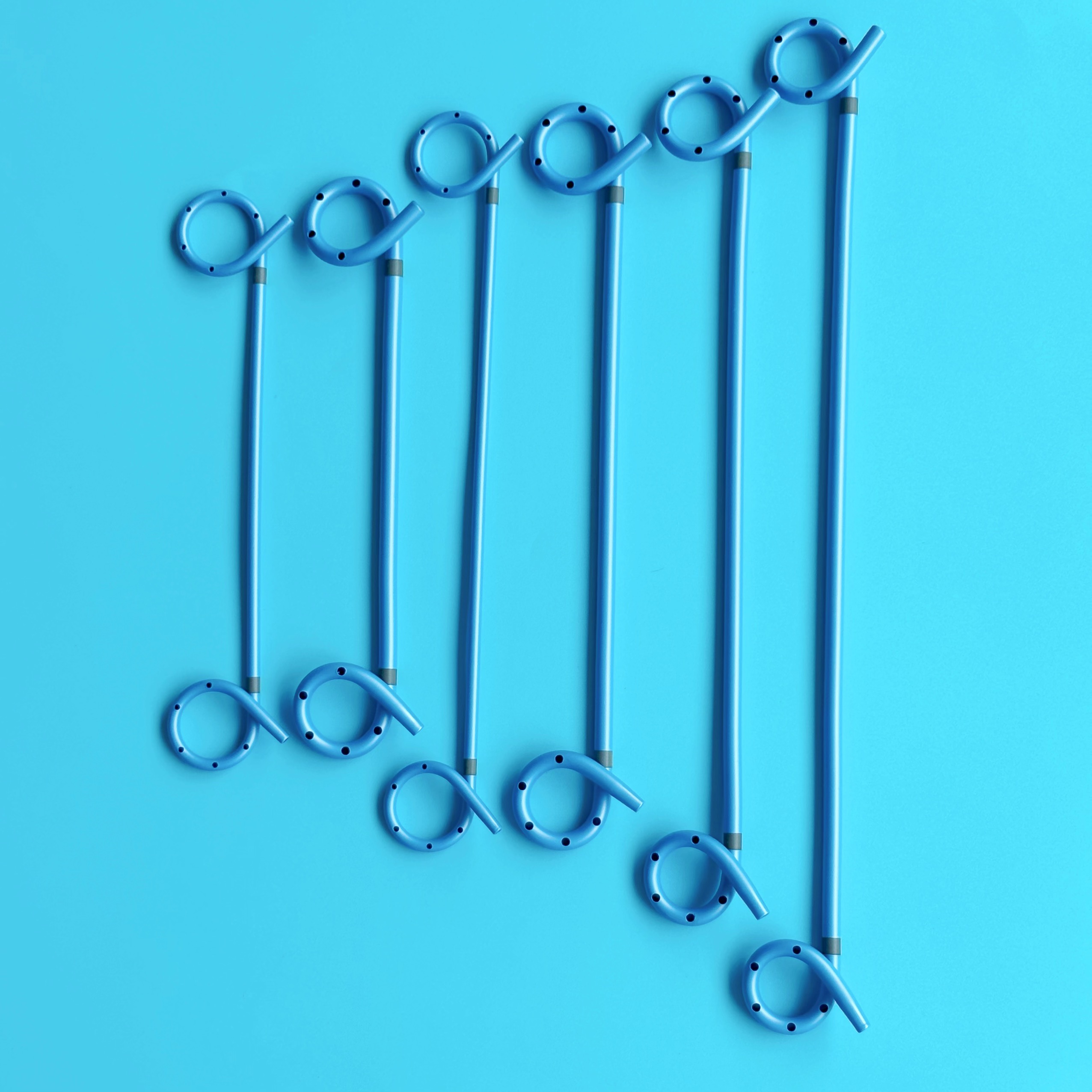
Clinical indications dictate the structural configuration. Understanding specific anatomical requirements helps avoid deployment errors. Selecting the correct loop format determines the long-term viability of the drainage tract.
The single pigtail drainage tube serves distinct functions. Primary use cases include percutaneous nephrostomy, biliary drainage, and pleural effusions. We also utilize it for localized abscesses. This device excels at internal-to-external drainage. When evaluating this option, focus heavily on external fixation points. Ensure secure connections to drainage bags or flow valves. You must carefully monitor the external exit site.
Internal stenting requires different mechanics entirely. The double pigtail drainage tube operates completely inside the body. Primary use cases include ureteral stents moving fluid from kidney to bladder. It also works perfectly for internal biliary drainage. Here, the evaluation lens shifts. Focus on dual-anchoring capabilities. One loop anchors the proximal end. The other secures the distal end. This dual-coil design effectively prevents migration within internal pathways.
Medical polymers have evolved rapidly over the last decade. Thermoplastic Polyurethane (TPU) stands out in the clinical context. TPU is an advanced elastomer blending extreme flexibility and robust mechanical strength. This makes it highly desirable for internal medical applications.
When compared to older materials like silicone or PVC, a TPU pigtail drainage tube offers notable performance benefits:
Kink Resistance: TPU maintains a wide internal lumen even when bent. This structural integrity reduces occlusion risks during patient ambulation.
Thermosensitivity: The material softens upon introduction to core body temperature. This unique property decreases internal tissue irritation. It directly improves overall patient comfort.
Tensile Strength: High tensile strength allows for thinner catheter walls. You maximize internal flow rates without increasing the external French size. This means a smaller puncture hole for the patient.
Clinical consensus now favors smaller interventions. Extensive literature demonstrates strong pleural drainage efficacy for coiled catheters. Studies show 8F–14F pigtail catheters yield comparable resolution rates for pneumothorax. They also perform exceptionally well for non-viscous effusions. They match the success rates of massive 24F+ chest tubes.
Furthermore, complication rates drop noticeably. Evidence highlights a reduced incidence of insertion-site pain. Organ puncture risks decrease sharply. We also see fewer cases of intercostal nerve damage. Smaller insertion tracks heal much faster and leave smaller scars.
However, we must operate under transparent assumptions. Clinical limitations still exist. Highly viscous empyema presents a challenge. Massive hemothorax or fluid containing heavy particulate matter might block smaller lumens. In those specific scenarios, patients may still require larger traditional tubes. You must assess fluid viscosity before device selection.
Feature | Pigtail Catheter (8F-14F) | Large-Bore Tube (24F+) |
|---|---|---|
Insertion Profile | Minimally invasive, Seldinger technique | Invasive, blunt dissection required |
Pain Level | Low to moderate | High, often requires heavy analgesia |
Fluid Type Suitability | Air, serous fluid, thin pus | Thick pus, clotted blood, heavy particulates |
Tissue Trauma Risk | Low (curved tip protects organs) | Moderate to High (straight tip) |
Even the best devices require vigilant management. Complications can arise if standard protocols slip. Anticipating these issues prevents clinical emergencies.
The risk of occlusion demands constant attention. Fibrin and cellular debris can accumulate inside the lumen. You must implement practical steps for managing catheter blockages. Establish routine sterile saline flushing protocols. Use fibrinolytics where clinically indicated to dissolve thick deposits. Regular flushing maintains the critical inner diameter.
Insertion challenges also require targeted training. The over-the-wire (Seldinger) technique represents the standard of care. However, it involves a learning curve. Clinicians must avoid wire kinking during insertion. A kinked guide wire makes catheter advancement impossible. It can also damage the TPU walls from the inside.
Patient monitoring serves as the final safeguard. Bedside staff must identify warning signs early. A sudden drop in drainage volume suggests positional occlusion or tubing kinks. Fluid leakage at the insertion site often indicates a blockage downstream. Promptly investigating these signs prevents severe drainage failure.
Selecting the correct device requires matching features to intended clinical outcomes. Use clear feature-to-outcome mapping. First, evaluate the viscosity of the target fluid. This directly informs the appropriate French size. For example, use 8F for air. Select 10-14F for serous fluid. Reserve 14F+ for thicker pus. Second, assess the duration of placement. Long-term drainage strongly dictates the necessity of a locking mechanism.
Compliance and manufacturing standards form the second review layer. Buyers must closely examine supplier documentation. Look for:
ISO Certifications: Ensures rigorous quality control during the manufacturing process.
Biocompatibility Testing Data: Confirms the material will not trigger systemic immune responses.
Radiopacity: Guarantees clear visibility under fluoroscopic or X-ray guidance for accurate placement.
Transitioning to specialized coiled catheters improves retention and patient comfort. It achieves this without sacrificing overall drainage efficiency. The memory-shaped loop drastically reduces accidental dislodgment.
Evaluators must utilize a robust final decision matrix. Always balance the material advantages of TPU against clinical needs. Select the optimal configuration between single and double loops based on anatomical pathways. Finally, determine if locking features align with your specific clinical caseload.
Your next steps should involve hands-on evaluation. We encourage clinical leads to request detailed product specification sheets. Obtain clinical evaluation samples for your team. Consult with a device specialist to drive institutional standardization and improve patient outcomes.
A: Typical dwell times range from a few days to several weeks. Safety depends heavily on the material and fluid type. TPU and silicone resist degradation better than PVC. However, prolonged placement increases the risk of encrustation and biofilm formation. Clinicians should routinely assess the patient and remove the catheter as soon as drainage resolves to prevent infection.
A: Yes, if properly sized. Larger French pigtails (12F to 14F) handle moderately viscous fluid and purulent drainage effectively. However, extremely thick fluids, heavy blood clots, or dense particulate matter often cause blockages. In those extreme cases, alternative management using larger traditional tubes or surgical washout remains necessary.
A: Several factors disrupt flow. The most common causes include positional occlusion, where the side ports rest flat against a cavity wall. Fibrin, thick pus, or blood clots can also physically block the internal lumen. Sometimes, a sudden stop simply means complete resolution of the fluid collection. Always flush the line to diagnose the exact issue.
A: Safe removal requires careful unlocking. You must unlock or cut the internal retention string at the hub before withdrawal. This releases the tension, allowing the coiled tip to straighten out. Pulling a locked catheter without releasing the string will cause severe tissue damage, bleeding, and immense pain for the patient.


