

close
Choose Your Site
Global
Social Media
Views: 0 Author: Site Editor Publish Time: 2026-06-26 Origin: Site


Uncontrolled fluid accumulation in pleural, biliary, or pericardial cavities presents serious clinical challenges. Rapid or unmanaged evacuation causes severe tissue trauma. It increases the risk of accidental dislodgement and sudden catheter occlusion. These adverse events threaten patient stability. They also complicate the overall recovery process significantly.
The single pigtail drainage tube serves as a highly reliable standard-of-care solution. Clinicians use it to manage complex fluid dynamics safely. It carefully balances essential patient comfort and consistent fluid evacuation. The distinctive curled geometry anchors the device securely. Simultaneously, it prevents the active suction holes from gripping delicate tissues.
We designed this article for clinical decision-makers and medical procurement teams. You will discover an objective framework for evaluating these critical devices. We explore specific design mechanics, advanced material science, and robust clinical safety profiles. You can use these insights to optimize patient care protocols across your departments.
Mechanism of Action: The coiled "pigtail" tip naturally anchors the tube and prevents the distal drainage holes from suctioning against adjacent tissue, ensuring continuous, controlled flow.
Material Matters: Thermoplastic Polyurethane (TPU) is the preferred standard, offering optimal kink-resistance and biocompatibility for prolonged indwelling times.
Versatile Efficacy: Single pigtail designs are highly adaptable, serving effectively in pleural drainage, abscess drainage, and as a biliary stent drainage tube.
Procurement Logic: Evaluating these devices requires assessing radiopacity, guidewire compatibility, locking mechanisms, and regulatory compliance (e.g., ISO, CE, FDA).
The coiled distal tip acts as a crucial physical barrier. It prevents active suction eyes from adhering directly to cavity walls. For example, it protects delicate structures like the pleura or peritoneum. This spatial buffer reduces localized patient pain significantly. It also minimizes unnecessary tissue trauma during continuous therapy. Clinicians observe far fewer complications related to mucosal damage.
Patient movement frequently dislodges traditional straight tubes. This creates emergency re-insertion scenarios. The pigtail shape functions as a low-profile internal anchor. Once deployed inside the cavity, the loop secures the device naturally. It drastically lowers the risk of accidental removal. The design requires less external traction overall. You can rely on its inherent structural stability during prolonged treatments.
Debris and fibrin often build up near standard catheter tips. This mixture quickly blocks conventional straight tubes. The multi-port pigtail design actively solves this problem. It places strategically spaced side holes along the protected inner curve. These inner holes optimize continuous fluid intake from multiple angles. The predictable flow prevents sudden system clogs. Your patients receive a steady, highly controlled drainage rate.
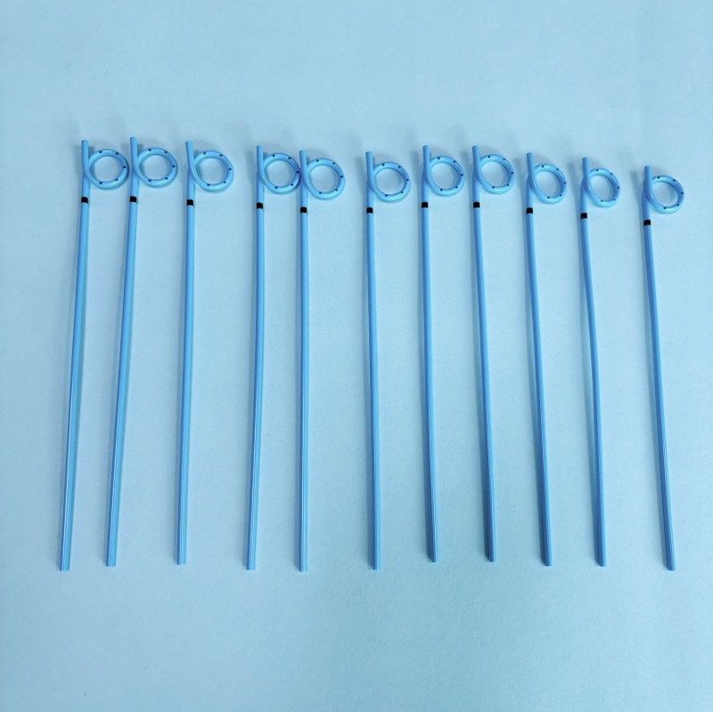
Comparing raw materials reveals clear clinical differences. Thermoplastic Polyurethane (TPU) dominates the modern device market. A reliable medical drainage catheter requires optimal tensile strength. TPU outperforms traditional silicone and older PVC compounds easily. Superior strength allows manufacturers to engineer much thinner walls. Thinner walls create a substantially larger inner lumen. This maximizes overall flow capacity effectively. Crucially, manufacturers do not need to increase the outer French size. Patients experience significantly less pain during insertion.
The advanced TPU drainage tube softens naturally at core body temperature. High-quality TPU reacts to body heat within minutes of placement. This thermosensitive softening improves the implementation reality dramatically. It reduces mechanical irritation at the dermal insertion site. Patients feel greater comfort during regular respiratory movements. They can safely mobilize earlier in their recovery.
Structural memory prevents dangerous fluid build-up. TPU demonstrates exceptional resistance to severe kinking when bent. The material bends with patient movement but refuses to collapse. This feature remains a critical safety requirement. It prevents sudden blockages and dangerous cavity pressure spikes. You maintain uninterrupted patency even in difficult anatomical positions.
Material Type | Tensile Strength | Kink-Resistance | Patient Comfort (In-Situ) |
|---|---|---|---|
TPU (Polyurethane) | Excellent (Allows thinner walls) | Very High | High (Thermosensitive softening) |
Silicone | Low (Requires thicker walls) | Moderate | High (Inherently soft) |
PVC | Moderate | Low to Moderate | Low (Rigid, causes irritation) |
Physicians trust these precise devices for thoracic interventions. They serve as safe alternatives to traumatic large-bore chest tubes. Clinicians use them for primary pneumothorax management. They also resolve malignant pleural effusions effectively. The standard Seldinger technique ensures minimally invasive placement. We recommend following these standardized procedural steps:
Identify the optimal insertion site using real-time ultrasound guidance.
Administer local anesthesia to minimize procedural discomfort.
Insert the introductory needle precisely into the target cavity.
Thread the compatible guidewire securely through the needle hub.
Advance the tapered tissue dilators over the guidewire gently.
Deploy the catheter, remove the wire, and engage the pigtail lock.
The specialized biliary stent drainage tube excels in complex gastrointestinal anatomies. Gastroenterologists use it routinely to bypass difficult biliary strictures. Interventional radiologists decompress the obstructed biliary tree safely. The internal pigtail secures the distal end firmly. It anchors well in the duodenum or the upper biliary tract. Clinicians can establish controlled external drainage immediately. It also supports highly effective internal-external fluid management.
Targeted abscess fluid collection requires prolonged clinical patience. Percutaneous drainage tackles deep intra-abdominal or pelvic abscesses well. Medical providers rely heavily on steady, gravity-assisted flow. Low-suction methods also perform exceptionally in these scenarios. These treatments often span several consecutive days or weeks. The coiled design maintains vital patency throughout the entire duration.
Managing occlusion risks remains a daily clinical priority. Any indwelling catheter can clog over extended timeframes. Highly viscous fluids or blood clots accelerate this process. Standard flushing protocols prevent complete system blockages. You must adhere to routine maintenance. We suggest adopting these specific practices:
Prepare a sterile syringe filled with normal saline.
Clean the catheter access port meticulously before attachment.
Inject the saline using slow, exceptionally gentle pressure.
Stop immediately if you encounter high mechanical resistance.
Document the fluid output volume and color accurately.
External securing practices prevent catastrophic dislodgement. The internal loop secures the deep distal tip. However, you must actively lock the external segment too. Use modern StatLock devices or robust suture wings. Proper external securing prevents unwanted tube migration. It also limits bacterial infection risks at the dermal entry point.
Common Mistake: Relying solely on the internal pigtail for full device stability. Always secure the external hub firmly to the patient's skin.
Device removal considerations require careful, deliberate attention. You cannot pull a locked pigtail abruptly. Engage the specific uncoiling mechanism prior to extraction. Most modern designs feature a dedicated unlocking string at the hub. You must release this tension string completely. Ensure the deep tip straightens fully inside the cavity. Proceed with smooth extraction to prevent painful tissue tearing.
Excellent radiopacity ensures precise placement verification immediately. Premium manufacturers integrate Barium Sulfate directly into the tubing matrix. This specific additive makes the device highly visible. Clinicians track it easily under live fluoroscopy. Standard X-ray and ultrasound imaging confirm correct positioning quickly. You must verify marker density before finalizing purchasing decisions.
Seamless guidewire and dilator compatibility speeds up critical workflows. Packaged catheter kits must match standard institutional guidewires perfectly. Verify exact fit for 0.035-inch or 0.038-inch wires. Smoothly tapered dilators reduce insertion friction significantly. They prevent painful dermal tearing during the initial placement phase.
The locking mechanism reliability dictates ongoing patient safety. Evaluate the specific string-lock or tension-lock mechanism thoroughly. It must remain intuitive for busy, stressed clinicians. Nursing staff should engage it securely in mere seconds. They must also disengage it effortlessly during final removal. Overly complicated locks cause unnecessary procedural delays.
Strict compliance and manufacturing standards mitigate serious institutional risks. Procurement teams must demand valid, up-to-date quality certifications. Verify ISO 13485 certification for medical quality management systems. Confirm comprehensive biocompatibility testing under the ISO 10993 framework. You should request proof of relevant regional regulatory clearances. CE marks and FDA approvals validate essential product safety.
Procurement Feature | Primary Clinical Benefit | Verification Method |
|---|---|---|
Barium Sulfate Integration | Allows precise anatomical positioning. | Test under standard X-ray/Fluoroscopy. |
0.035" / 0.038" Compatibility | Prevents delays during Seldinger placement. | Review manufacturer kit specifications. |
Tension-Lock Hub | Ensures the internal loop remains formed. | Conduct physical hands-on staff trials. |
ISO 10993 Certification | Guarantees long-term tissue biocompatibility. | Request official compliance documentation. |
We have reviewed the core mechanics driving successful fluid evacuation. A highly engineered geometry fundamentally transforms fluid management protocols. The unique looped design actively prevents unpredictable fluid accumulation. It anchors firmly while protecting adjacent sensitive tissues. Premium materials like advanced TPU guarantee long-term patient safety.
Facilities can finally standardize their controlled drainage protocols confidently. We recommend taking immediate, actionable steps. Clinical directors should request physical sample kits for hands-on evaluation. Test the radiopacity under your own institutional imaging equipment. Verify direct compatibility across your existing drainage bags. Implement these standardized checks to elevate your departmental patient care metrics.
A: A single pigtail has one coiled end (usually placed internally) and straightens externally for connection to a drainage bag. A double pigtail has coils at both ends, typically used for internal stenting (e.g., ureteral or internal biliary stenting) to prevent migration in either direction.
A: Indwelling time varies by clinical indication and regulatory approval, but high-grade TPU is highly biocompatible. It is typically approved for up to 29 days, provided regular site care and patency checks are maintained.
A: Yes, they can be connected to low-pressure active suction systems or gravity drainage bags. However, the exact suction pressure should be dictated by clinical guidelines specific to the fluid cavity (e.g., pleural vs. biliary).
A: Many pigtail catheters feature a tension string running through the lumen. Once the catheter is in place, the clinician pulls and locks the string at the hub, which forcefully forms and maintains the pigtail loop inside the cavity.


