

close
Choose Your Site
Global
Social Media
Views: 0 Author: Site Editor Publish Time: 2026-03-16 Origin: Site


After surgery, many patients ask: how long does a drainage tube stay in? A surgical drainage tube removes fluid and supports healing. The drainage tube removal time depends on surgery type and daily drainage volume. In this article, you will learn drainage tube duration and removal timing. Reliable drainage tube products from Bova help maintain stable fluid drainage. Learn more about our products.
How Long Does a Drainage Tube Stay In After Surgery?
Most surgical drainage tubes stay in place for about 7 to 14 days. This period allows the body to release excess blood, lymph fluid, or inflammatory fluid produced during healing.At the beginning, drainage volume is usually higher. As tissues recover and swelling decreases, the amount of fluid gradually drops.Doctors monitor this change carefully. When fluid production becomes minimal, the drainage tube can usually be removed safely.
Drainage tube removal time is rarely determined by days alone. Instead, surgeons evaluate the amount of fluid collected each day.If drainage output remains high, removing the tube too early could allow fluid to accumulate again. This buildup may cause swelling, infection, or delayed healing.Therefore, surgeons wait until fluid output falls below a safe threshold before removing the tube.
In many surgical cases, doctors consider removing the drainage tube when daily output drops below 20–30 milliliters within 24 hours.However, this threshold may vary depending on the surgery type and the patient's condition.
Note: Each hospital or surgeon may use slightly different guidelines for drainage tube removal.
Certain surgeries produce larger fluid volumes. In these situations, drainage tubes may stay in place longer.For example, cancer surgeries, reconstructive procedures, or orthopedic operations often require longer drainage periods.Patient healing speed also affects drainage tube duration.
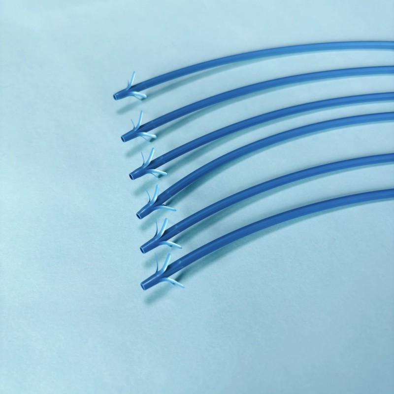
Large surgical procedures often create more tissue trauma. This damage activates the body’s inflammatory response and increases fluid production during healing. As a result, doctors often place a surgical drainage tube to prevent fluid accumulation and reduce pressure around the surgical site.
Operations such as joint replacement, abdominal surgery, or cancer tumor removal usually involve deeper tissue layers. These procedures tend to produce more postoperative drainage. Because the body needs more time to clear blood, lymph fluid, and inflammatory fluid, the drainage tube may remain in place longer.
In contrast, smaller procedures usually generate less fluid. Minor soft tissue surgeries or small incision procedures may require drainage for only a few days. Surgeons consider the procedure type when estimating drainage tube duration.
Every patient heals at a different pace. Age, immune health, and nutrition all influence tissue repair speed. When healing is faster, the body produces less excess fluid and drainage levels decrease sooner.Patients with strong immune systems often show faster wound healing. Their drainage volume drops quickly, allowing doctors to remove the drainage tube earlier.
Older patients or individuals with chronic illness may recover more slowly. Conditions such as diabetes or cardiovascular disease can delay tissue repair. In these cases, fluid production may continue longer and drainage tubes may remain in place for extended periods.
Doctors measure drainage output every day. This is one of the most important indicators used to determine when a drainage tube can be removed safely.At the beginning of recovery, drainage volume is usually higher because the body releases blood and inflammatory fluid from the surgical area. As healing progresses, the amount gradually decreases.When daily drainage falls below a safe threshold, often around 20–30 milliliters within 24 hours, doctors may decide to remove the tube.
Surgeons sometimes keep drainage tubes in place longer to prevent complications. These may include bleeding, infection, or fluid buildup around the wound.Keeping the drainage tube temporarily allows doctors to monitor the surgical site and continuously remove fluid. This reduces pressure inside the tissue and lowers infection risk.Removing the tube too early may allow fluid to collect under the skin or inside the body cavity. This can cause swelling, delayed healing, or additional procedures.
Different drainage systems are selected based on surgical location, expected fluid volume, and the type of tissue involved. Each system functions differently and may remain in place for a different period.
The Jackson-Pratt drain is one of the most widely used surgical drainage systems. It consists of a flexible tube connected to a small suction bulb. When the bulb is compressed, it creates gentle negative pressure that pulls fluid away from the surgical area.Because the system is compact and portable, patients often go home with the drain still in place and empty the bulb themselves.Jackson-Pratt drains usually remain in place for one to two weeks, depending on daily drainage levels.
Hemovac drains use a spring-loaded suction reservoir instead of a bulb. This design allows the system to collect a larger volume of fluid.These drains are commonly used after orthopedic procedures such as joint replacement or spinal surgery. These operations often generate significant postoperative drainage.Hemovac drains typically stay in place several days to about one week, depending on how quickly drainage decreases.
Penrose drains rely on gravity rather than suction. The open tube allows fluid to flow naturally from the wound into a dressing.They are frequently used in infected wounds or abscess drainage because they allow thick fluids and pus to exit easily.Penrose drains usually remain in place for a few days, until the infection fluid drains adequately.
Chest tubes are specialized drainage systems used in thoracic surgeries. They remove air, blood, or fluid from the chest cavity after lung or heart procedures.These tubes help the lungs expand properly and prevent pressure buildup inside the chest.Chest tubes remain in place until air leakage stops and fluid drainage decreases significantly. This process may take several days or longer depending on the surgical condition.
Drain Type | Drainage Method | Typical Duration |
Jackson-Pratt | Suction bulb | 7–14 days |
Hemovac | Vacuum suction | 3–7 days |
Penrose | Gravity drainage | 2–5 days |
Chest tube | Controlled chest drainage | Several days |
Tip: Hospitals usually select the drainage system based on expected fluid volume and surgical location. Choosing the correct system helps improve recovery efficiency.
The most reliable sign that a drainage tube can be removed is a consistent decrease in drainage volume.When daily drainage becomes minimal and remains stable for several days, it indicates that the body no longer requires active fluid removal.
Drainage fluid often changes color during healing.At the beginning, the fluid may appear dark red because it contains blood from the surgical site. As healing continues, it usually turns pink and eventually becomes light yellow or clear.This color transition indicates normal wound healing.
As excess fluid leaves the body, swelling around the surgical area decreases.Patients often notice reduced tension or tightness near the incision site. This indicates that fluid accumulation inside the tissue is declining.
Doctors also evaluate overall recovery progress. They check for signs of infection, abnormal bleeding, or increasing pain.If the patient's condition remains stable and healing progresses normally, drainage tube removal becomes safe.
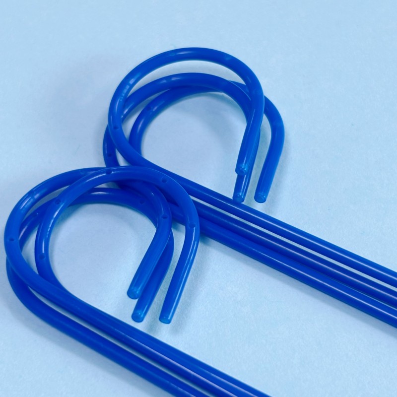
Healthcare providers measure drainage output regularly. This data helps determine whether the drainage tube is still necessary.If drainage levels remain high, the tube stays in place longer. Once fluid output decreases steadily, removal becomes possible.
Drainage tubes are usually removed during scheduled follow-up appointments.During the visit, the surgeon examines the wound and reviews recorded drainage levels to determine if removal is appropriate.
Every patient’s situation is unique. Surgeons evaluate the type of surgery, healing progress, and risk of complications.Based on these factors, they decide the safest time to remove the drainage tube.
Note: Surgeons avoid removing the tube too early because fluid buildup inside the wound can delay healing and increase infection risk.
Removing a drainage tube is usually a quick and straightforward procedure. The doctor first removes the stitch holding the tube in place.The tube is then gently pulled out from the surgical site.
Most patients describe the removal process as slightly uncomfortable but not painful.The entire procedure usually takes only a few seconds.
After removal, the doctor places a sterile dressing over the small opening where the tube was located.The opening normally closes naturally within a few days.
Patients should keep the wound area clean and dry.Monitoring the site for redness, swelling, or unusual drainage is important during the early healing period.
Proper hygiene helps prevent infection. Patients should gently clean the area around the drainage tube using soap and water.Keeping the insertion site dry and clean reduces the risk of bacterial contamination.
Most drainage systems include a collection bulb or container. Patients often need to empty it several times per day.Doctors may ask patients to record the drainage amount and fluid color to monitor healing.
The drainage tube should be secured to clothing or bandages to prevent pulling on the skin.Loose clothing can also help reduce pressure on the drainage site.
Patients should monitor for symptoms such as fever, swelling, foul odor, or sudden drainage changes.These signs may indicate infection or complications and should be reported to a doctor immediately.
Tip: Always wash hands before and after handling the drainage system to reduce infection risk.
Observation | Normal Sign | Warning Sign |
Drainage color | Light pink or yellow | Dark red or cloudy |
Drainage amount | Gradually decreasing | Sudden increase |
Tube position | Secure and stable | Tube displacement |
Some surgical procedures naturally produce fluid for longer periods. In these cases, the drainage tube must remain in place until fluid output decreases.This prevents fluid accumulation that could slow healing.
Sometimes thick fluid or tissue debris can block the tube. When this happens, drainage may stop temporarily.Doctors may flush or adjust the drainage tube to restore normal fluid flow.
If infection develops, fluid production may increase and swelling may appear around the drainage site.Doctors may keep the drainage tube in place longer while treating the infection.
Occasionally, internal bleeding may occur after surgery. This increases drainage volume and requires close monitoring.Doctors must identify and control the bleeding before removing the drainage tube safely.
Problem | Cause | Solution |
Tube blockage | Thick drainage fluid | Tube flushing |
Infection | Poor hygiene | Antibiotic treatment |
Fluid buildup | Early removal | Reinsertion if needed |
A drainage tube usually stays in place for several days to two weeks after surgery. Doctors monitor drainage levels and healing before removal. Stable drainage supports safer recovery. High-quality drainage tube systems from Bova offer multi-hole drainage, anti-kink tubing, and clear positioning for reliable postoperative fluid management.
A: A drainage tube usually stays in for several days to about two weeks, depending on postoperative drainage and healing progress.
A: A surgical drainage tube is removed when daily drainage falls to a low level and wound healing is stable.
A: A drainage tube stays in the body to remove fluid, prevent swelling, and reduce infection risk after surgery.
A: Drainage tube removal time depends on surgery type, daily drainage volume, and the patient’s recovery speed.


